"If all the matter in the universe except the nematodes were swept away, our world would be still be dimly recognizable, and if, as disembodied spirits, we would investigate it, we should find its mountains, hills, vales, rivers, lakes and oceans represented by a thin layer of nematodes..."
- N. A. Cobb, nematologist
Free living nematodes:
http://www.ucmp.berkeley.edu/phyla/ecdysozoa/nematoda.html
There are 20,000 described species, although conservative estimates promise 500,000 to 1 million spp. in the near future. They occupy a wide range of habitats including beer-soaked mats in pubs and permafrost soils in Arctic and Antarctic tundra.

Nematode Characteristics
Collagenous (protein) cuticle with many layers, that must be shed to grow.
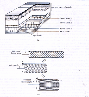
It is flexible, but not elastic and can serve as an antagonist when thick enough to longitudinal muscles. It enable nematodes to maintain high internal pressures, it is not uncommon for pressures equal to 70mm of mercury, with a maximum recorded value of 125mm of mercury. The cuticle bears 4 sensory ridges internally. Fibers within the cuticle are arranged at different angles to each other creating a meshwork of reinforcing strands that strengthen the body wall against hydrostatic pressure, and at the same time, permit the cuticle to bend.

The body cavity is derived from blastocoel and is a pseudocoelom. The pseudocoelom is loosely filled with tissue, or fluid-filled, and used in combination with longitudinal muscles to create a hydrostatic skeleton. Without internal divisions in the pseudocoelom, contraction of any of the muscles affects the entire length of the animal causing sinusoidal, whiplike movements as muscle on different sides of the body contract.
Body is thin, elongated, cylindrical, with both ends tapered. Three to six labia surround mouth and bear sensillae. Buccal cavity may have styles or teeth
A long muscular pharynx, and triangular intestinal lumen that ends in terminal anus. Because of the constant hydrostatic pressure on the pseudocoelom, nematodes have a system of circular muscles forming valves in the pharynx so they can swallow food. When the posterior valve closes, the anterior opens and the pharynx sucks in food.
The Brain is circular around pharynx (Cycloneuralia).
We will be placing a number of small phyla in this group.
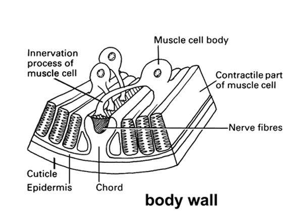
Muscles branch to dorsal and ventral nerve cords, rather than nerves to muscles.

Lateral cords contain excretory tubes and smaller nerves. Nematodes have no circulatory or respiratory organs and the excretion of metabolic waste is via two simple ducts or tubules which have no nephridia or flame cells.
Reproduction
As a whole the phylum is characterized by species with separate sexes, but a few species may be parthenogenetic or hermaphroditic. For example, in C. elegans, the hermaphrodite is XX and the male is XO. Sex is determined by the ratio of X chromosomes to autosomes. In closely related species of nematodes, XX females are found, suggesting that the hermaphrodites evolved from females. Somatically, the females and hermaphrodites are identical, the only difference being that the hermaphrodites make sperm during their early development before switching over to egg production. In C. elegans, there even exists a dominant mutation (tra-1D) that transforms XX or XO individuals into fertile females. In colonies with such an allele, three sexes are possible and functioning (Hodgkin, 1980).
Females are generally larger than males. Fertilization is internal. Copulatory spicules hold the male in place as he passes amoeboid sperm to the female.
Cleavage is non-spiral and involves strange chromosome diminution in non germ cells. Chromatin diminution is defined as chromosomal fragmentation, followed by the elimination of part of the chromosome during mitosis. By carefully analyzing the cell lineage, Boveri (1910)demonstrated that all cells which undergo chromatin diminution, and therefore contain less chromatin, became somatic cells, whereas nuclei retaining the original integrity of the chromosomes and the full quantity of chromatin gave rise to germ line cells . Thus the process of chromatin diminution is clearly linked to germ line-soma differentiation and in fact serves to distinguish cytologically the germ line and the somatic cell lineages.
Encyclopedia of Life Sciences
Published by John Wiley & Sons, Ltd
Figure 1. Chromatin diminution in Parascaris univalens. (a) Anaphase of the first cleavage division. (b) Anaphase of the second cleavage division. Chromatin diminution occurs in the upper S1 cell but not in the lower P1 cell. (c) Four-cell stage after completion of the second cleavage division. The cells S1a, S1b and S2 give rise to the somatic cells, while the P2 cell represents the germ line. P0, zygote; C, centromere; E, eliminated chromatin; N, nucleus; P0-P2, germline; S1, S1a, S1b, S2, presomatic cells. From Müller et al. (1996).
Development is strongly determinate and in many species except in the germ cells, there is a fixed number of cells, or eutely.
The Blastopore becomes mouth. Individuals molt through 4 juvenile instars (ecdysis) and resemble adults, but are called larvae. Use to be given specific names, but now simply refered to as LI, L2, L3 and L4.
Many have the ability to withstand extremely dry, cold conditions, an adaptation known as cryptobiosis.
Economical and scientific importance
Nematodes are used as biological models. A free-living species with the scientific name C. elegans is widely used in genetics, the neurosciences, and developmental biology.
Examples:
Used to study chromosome dimunitation and so how this affects gene function.
Used to study the regulation of germ line/somatic line determination.
Because of their simplicity they have been used to study the genetics of most processes including the development of memory.
Encyclopedia of Life Sciences
Published by John Wiley & Sons, Ltd
Figure 2. Apparatus used to assess habituation in C. elegans. Individual worms are placed on to a Petri dish, held in a plastic attachment that is connected to a micromanipulator. A mechanical tapper driven by an electromagnetic relay is used to deliver the taps in a consistent and uniform manner to the Petri dish from a Grass S88 stimulator. A video camera attached to the microscope and connected to a video monitor and video cassette recorder is used to monitor and record the behavior of the worm. To allow for precision of the timing of events, a time–date generator is used to superimpose the time (in thousandths of a second) and date of the experiment onto both the monitor and the recorder. After habituation training, the magnitude of the responses to tap are quantified using stop-frame video analysis by tracing each response on to an acetate, which is then scanned into a microcomputer to measure the length of each response. (Adapted from Peters et al. 1999).
The genome of C. elegans has been completely sequenced.
They are also being exploited as pest control.
Hundreds of researchers representing more than forty countries are working to develop nematodes as biological insecticides. Nematodes are sold in the U.S., Europe, Japan, and China for control of insect pests in high-value horticulture, agriculture, home and garden niche markets.
Nematode exits from a spider.
Turning the tables on nematodes.
The nematode-trapping and endoparasitic fungi are found in all major taxonomic groups of fungi, and they occur in all sorts of soil environments where they survive mainly as saprophytes. The ability to use nematodes as an additional nutrient source provides them with a nutritional advantage. The fungi enter their parasitic stage when they change their morphology and traps or mature spores are formed. The trapping (predatory) fungi have developed sophisticated hyphal structures, such as hyphal nets, knobs, branches or rings, in which nematodes are captured by adhesion or mechanically. The endoparasites, on the other hand, attack nematodes with their spores, which either adhere to the surface of nematodes or are swallowed by them. Irrespective of the infection method, the result is always the same, the death of the nematode.
Some parastistes of concern for humans.
Ascaris lumbricoides is one of the best known of the parasitic nematodes. Males and females live in the intestines of humans where they graze on intestinal contents. Eggs pass out with the feces and, if they contaminate food, are introduced to another host. The larvae hatch out in the intestine of the new host and then burrow through the walls to be carried by the bloodstream to the lungs. At the lungs they burrow through the alveoli and crawl up the trachea and down the esophagus. Larvae usually burrow out of the lungs at night and are unknowingly swallowed by the host. Occasionally larvae get lost and crawl up the esophagus and exit at the nose. In some areas of the world Ascaris is so common that a child is not considered to be part of the tribe until a larva is sneezed out and found in the bed.http://www.cdc.gov/parasites/ascariasis/biology.html
"While preparing a routine sagittal section of the human head and neck for the teaching of anatomy, 3 mature Ascaris lumbricoides worms were seen in the maxillary sinus and 3 in the sphenoidal sinus. Of the worms in the maxillary sinus, 2 were males and one was female, while in the sphenoidal sinus all 3 worms were females. The worms measured 7 - 11 cm in length Light microscope examination of the maxillary sinus wall revealed a few eggs of the parasite in close relation to the epithelium. The migratory abilities of A. lumbricoides adult worms are well documented and most often involve the bile and pancreatic ducts (Asrat & Rogers, 1995). In a endemic area of Kashmir, India, A. lumbricoides was the cause of acute pancreatis in 23 percent of 256 patients (Khuroo et al., 1992). Worm extraction and biliary drainage were indicated in 32 percent of 156 patients who had acute hepatobiliary and pancreatic ascariasis, and this resulted in rapid relief of symptoms in most patients (Khuroo et al., 1993). In the case represented, the ostia of the sinuses were between 3 and 4 mm in width and were therefore large enough for the passage of the worms as adults."
A case of Ascaris suum bioterrorism occurred at a Canadian university in 1970, when a postgraduate student in parasitology contaminated the food of four roommates with worm eggs. All experienced symptoms of lower respiratory tract disease, and two of them developed acute respiratory failure.
Hooks are found in most domestics and can be a problem for humans: Necator and Ancyclostoma. http://www.cdc.gov/parasites/hookworm/biology.html
Adults live in intestine, feed on cellular lining, causing bleeding. Eggs are released in feces. Larvae hatch in soil, climb on grass, and penetrate bare skin of new host. They then migrate through bloodstream and out through intestine
Pinworm: Enterobius vermicularis is common in the South of the United States. http://www.cdc.gov/parasites/pinworm/gen_info/faqs.html
There are major differences in life cycle among nematode parasites.
|
|||||||
|
Eggs | Larva 1 | Larva 2 | Larva 3 | Larva 4 | Adults | |
|
Eggs | Larva 1 | Larva 2 | Larva 3 | Larva 4 | Adults | |
|
Eggs | Larva 1 | Larva 2 | Larva 3 | Larva 4 | Adults | |
|
Eggs | Larva 1 | Larva 2 | Larva 3 | Larva 4 | Adults | |
|
Microfilariae | Larva 1 | Larva 2 | Larva 3 | Larva 4 | Adults | |
Trichinosis Worm (Trichinella) http://www.cdc.gov/parasites/trichinellosis/gen_info/faqs.htmlSome species have two two hosts cycles: filarial nematodes
Elephantisis: Caused by filarial nematodes that reside in the lymph vessels. http://www.cdc.gov/parasites/lymphaticfilariasis/biology.html
The worm can infect any species of mammal that consumes its encysted larval stages. When an animal eats meat that contains infective Trichinella cysts, the acid in the stomach dissolves the hard covering of the cyst and releases the worms. The worms pass into the small intestine and, in 1–2 days, become mature. After mating, adult females produce larvae, which break through the intestinal wall and travel through the lymphatic system to the circulatory system to find a suitable cell. Larvae can penetrate any cell, but can only survive in skeletal muscle. Within a muscle cell, the worms curl up and direct the cell functioning much as a virus does. The cell is now called a nurse cell. Soon, a net of blood vessels surround the nurse cell, providing added nutrition for the larva insideThis worm is acquired by eating undercooked flesh of pigs or other omnivorous mammals. Larvae burrow into intestinal lining, mature, reproduce, and release offspring. Juveniles invade muscles and form cysts, causing extreme pain.
Guinea worm
Guinea worm disease is caused by Dracunculus medinensis, a threadlike parasitic worm that grows and matures in people. Worms grow up to 3 feet long and are as wide as a paper clip wire.
People get infected when they drink standing water containing a tiny water flea that is infected with the even tinier larvae of the Guinea worm. Over the course of a year in the human body, the immature worms pierce the intestinal wall, grow to adulthood, and mate. The males die, and the females make their way through the body, maturing to a length of as much as 3 feet, and ending up near the surface of the skin, usually in the lower limbs. The worms cause swelling and painful, burning blisters. To soothe the burning, sufferers tend to go into the water, where the blisters burst, allowing the worm to emerge and release a new generation of millions of larvae. In the water, the larvae are swallowed by small water fleas, and the cycle begins again.
The only treatment is to remove the worm over many weeks by winding it around a small stick and pulling it out a tiny bit at a time. Sometimes the worm can be pulled out completely within a few days, but the process usually takes weeks or months. http://www.cdc.gov/parasites/guineaworm/disease.html
Dog Heartworm
http://www.heartwormsociety.org/pet-owner-resources/canine.html
https://www.heartwormsociety.org/veterinary-resources/practice-tools/heartworm-image
Usually takes weeks or months. (Dirofilaria)
The adult lives in dog’s heart muscle and eggs hatch into tiny larvae (“microfilariae”), which disperse in blood. The microfilariae are picked up by mosquito that bites dog and the mosquito transfers them to new dog. They migrate to heart and grow and mature in heart muscle.
Parasitic nematodes especially hook worms have been called "primitive" parasites. Why?
Nematodes are able to undergo a process known as cryptobiosis where normal life processes and functions are suspended during periods of environmental instability and inhospitability. http://en.wikipedia.org/wiki/Cryptobiosis
"The author, after permitting a number of female pig Ascuris to become extremely dry, placed them at a temperature of from - 5" C. to 10" C. (in a refrigerator) for a
period of three years. Observations were made to determine the amount of evaporation that takes place when female Ascaris are subjected to the conditions described above, and
found that they will dry to within approximately 5 per cent
(4.92 per cent) of total desiccation by the end of two months.
After one year, an Ascaris was removed from this temperature
and placed in distilled water for one day. The uterus,
still in a dried condition, was removed and rubbed up in a
mortar in order to force out the eggs. The eggs were then
placed in an incubator at a temperature of from 31" C. to
34.5" C., and at the end of ten days fully developed embryos
were noted. At the end of 25 months another Ascaris was removed and fully developed embryos were found at the end of 25 months. "from studies on the Ascaris lumbriocoides 1926, Harry mathias Martin, http://extension.unl.edu/publications
So their survival strategies are almost as note worthy as those of tardigrades.
Fossil history
The oldest described nematode fossil is preserved inside its fly host in Lebanese amber from the Lower Cretaceous (120–135 million years ago). Amber (fossilized resin) has been found to contain the remains of insect parasitic, plant parasitic and free-living nematodes. Fossil nematodes associated with vertebrates, including humans also exist. Nematodes (strongyles) have been recovered from the intestines of Upper Pleistocene horses and encysted juveniles of Trichinella have been recovered from the Egyptian mummy, Nakht, dated at about 1200 BC. Nematodes have also been recovered from 3000-year-old human coprolites from Lovelock Cave in Nevada, USA. Nematodes as a group are undoubtedly much older than the oldest fossils indicate and probably evolved in the Cambrian or Precambrian with free-living micro forms that fed on bacteria in aquatic or semi-aquatic habitats.